
Other Possible Causes of White or Light Spots
IGH is only one possible explanation for hypopigmented skin. Several other conditions can cause white or lighter patches, and some require very different treatment.
Among the possibilities are tinea versicolor, vitiligo, and post-inflammatory hypopigmentation.
- Tinea Versicolor
Tinea versicolor, also known as pityriasis versicolor, is a common superficial fungal condition associated with an overgrowth of Malassezia, a type of yeast normally found on human skin.
Unlike the tiny discrete spots commonly associated with IGH, tinea versicolor frequently produces larger patches that may be lighter, darker, pinkish, tan, or brown depending on the person’s natural skin tone.
Typical locations include:
Upper chest
Upper back
Shoulders
Upper arms
Neck in some people
The affected skin may have a very fine scale. Some people experience mild itching, while others notice no discomfort at all.
The color difference can become especially noticeable after sun exposure because affected areas may not tan in the same way as surrounding skin.
How Is Tinea Versicolor Different From IGH?
Location and texture can provide clues. IGH is particularly common on the shins and forearms and is generally smooth. Tinea versicolor often affects the trunk and shoulders and may have a fine, powdery scale.
Tinea versicolor is also associated with yeast overgrowth and can respond to antifungal treatment, whereas antifungal medication would not be expected to treat IGH.
This is one reason it is better to establish the diagnosis before repeatedly applying medications to unexplained white spots.
Treatment for Tinea Versicolor
Depending on the extent of the condition, treatment may involve topical antifungal products. A clinician or pharmacist can advise on appropriate options, particularly when the diagnosis is uncertain or the condition is extensive or recurrent.
Even after the yeast has been successfully controlled, normal pigmentation may take time to return. Persistent color difference immediately after treatment therefore does not necessarily mean that treatment has failed.
- Vitiligo
Vitiligo is quite different from IGH. It is a condition in which melanocytes, the cells responsible for producing pigment, are lost from affected areas.
Vitiligo often produces clearly defined areas of depigmented skin. These patches can be much larger than typical IGH spots and may gradually expand or appear in additional areas.
Vitiligo can occur almost anywhere, including:
Hands and fingers
Face
Areas around the mouth and eyes
Elbows
Knees
Feet
Genital areas
Other parts of the body
Some patterns are symmetrical, meaning similar areas on both sides of the body are affected.
Why Does Vitiligo Develop?
Vitiligo is generally regarded as an autoimmune condition. In autoimmune diseases, the immune system mistakenly targets the body’s own cells or tissues. In vitiligo, pigment-producing melanocytes are affected.
Genetic susceptibility can play a role, and vitiligo sometimes occurs alongside other autoimmune conditions.
It is not contagious and cannot be transmitted by touching someone who has it.
Does Vitiligo Need Treatment?
Vitiligo does not always require treatment from a general health perspective, but people may choose treatment for cosmetic reasons or because of how the condition affects their quality of life.
Depending on the person’s age, location of the patches, extent of involvement, and other factors, dermatologists may discuss topical medications, light-based treatments, camouflage products, and other therapies.
Treatment decisions should be individualized because no single approach is appropriate for everyone.
- Post-Inflammatory Hypopigmentation
Sometimes skin becomes lighter after inflammation or injury. This is known as post-inflammatory hypopigmentation.
It can occur after:
Eczema
Dermatitis
Minor skin injuries
Certain infections
Other inflammatory skin conditions
A useful clue is that the lighter area often appears where a previous rash, injury, or inflammatory problem occurred.
The pigmentation may gradually improve as the skin recovers, although the time required varies considerably.
Why Does Inflammation Change Skin Color?
Inflammation can temporarily disrupt normal pigment production or distribution. The result may be an area that looks noticeably lighter than surrounding skin after the original rash has resolved.
This can be particularly noticeable in people with medium or darker skin tones because there is greater contrast between the affected and unaffected areas.
Protecting the area from excessive sun exposure can also reduce contrast while the pigmentation gradually normalizes.
- Other Causes of Hypopigmentation
There are additional causes of white or lighter skin that cannot reliably be distinguished through a simple checklist. Certain inflammatory disorders, infections, scars, medication-related changes, and less common dermatologic conditions can alter pigmentation.
That is why persistent or unusual changes are best assessed by a healthcare professional rather than diagnosed exclusively from online photographs.
How Can You Tell IGH, Tinea Versicolor, and Vitiligo Apart?
There is some overlap, but several general characteristics can provide useful clues.
IGH: Often consists of numerous tiny, smooth white spots, particularly on sun-exposed arms and legs, and becomes more common with age.
Tinea versicolor: Often causes larger or merging patches on the chest, back, shoulders, or upper arms and may have fine scaling.
Vitiligo: Usually produces more clearly depigmented patches that can be larger, may expand, and can occur almost anywhere.
Post-inflammatory hypopigmentation: Often follows a previous rash, eczema flare, injury, or other inflammation in approximately the same area.
These descriptions are helpful for understanding the possibilities, but they should not be used as a substitute for diagnosis.
Does the Size of the Spots Matter?
Size can be a useful clue. Classic IGH lesions are generally very small. A large area of pigment loss, particularly one that continues expanding, is less typical and deserves evaluation.
However, size alone cannot determine the cause. Doctors consider the complete pattern, including the number of spots, location, borders, texture, symptoms, duration, and changes over time.
Does It Matter Whether the Spots Are Scaly?
Yes. Texture can provide useful information.
IGH is usually smooth. Vitiligo is also generally smooth because it primarily involves pigmentation rather than producing a surface scale.
A fine scale, especially on patches across the chest, shoulders, or back, may suggest a different process such as tinea versicolor or another inflammatory condition.
Persistent scaling, itching, redness, cracking, or inflammation should therefore be mentioned to a healthcare professional.
Why White Spots Can Look Worse in Summer
People frequently report that white spots become much more obvious during summer.
Sometimes the spots themselves have not changed significantly. Instead, the surrounding normal skin becomes darker after ultraviolet exposure, increasing the contrast.
This can happen with several forms of hypopigmentation.
Using sunscreen can reduce tanning of the surrounding skin and may make the contrast less dramatic while also protecting against ultraviolet damage.
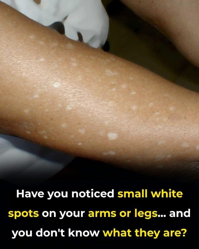
Once you understand what may be causing the spots, the next question is whether anything can actually prevent new ones or make existing ones less noticeable.You may notice them while getting dressed, standing in bright sunlight, or looking closely at your arms or legs: tiny white spots scattered across otherwise normal-looking skin. They may be completely flat, painless, and easy to ignore at first. Over time, however, you might begin noticing more of them and wonder whether they are simply part of aging or a sign that something is wrong.
Small areas of lighter skin are surprisingly common. In many adults, particularly those who have accumulated years of sun exposure, one possible explanation is a benign condition called idiopathic guttate hypomelanosis, usually abbreviated as IGH.
However, not every white spot is IGH. Fungal skin conditions, vitiligo, previous inflammation, and several other causes can also produce lighter areas of skin. Appearance alone is not always enough to make a reliable diagnosis.
The good news is that many causes of small white spots are harmless or manageable. Understanding their size, location, texture, pattern, and how they change over time can help you know when the spots are likely to be benign and when it makes sense to have them evaluated by a healthcare professional.
What Are Those Tiny White Spots?
Skin color is largely influenced by a pigment called melanin. Melanin is produced by specialized cells called melanocytes. When an area of skin contains less pigment than the surrounding skin, it can appear lighter or white.
This change is generally described as hypopigmentation or depigmentation, depending on how much pigment has been lost. There are numerous possible causes, and they do not all behave in the same way.
For example, a few tiny smooth white dots on the shins of an older adult may have a very different explanation from a large white patch that has gradually expanded over several months.
That distinction is important because the phrase “white spots” describes an appearance rather than a diagnosis.
The Common Possibility: Idiopathic Guttate Hypomelanosis
Idiopathic guttate hypomelanosis is a benign pigmentation condition characterized by small areas of lighter or white skin. It is commonly noticed on parts of the body that receive substantial sun exposure, particularly the arms and lower legs.
The medical name sounds complicated, but each part helps describe the condition.
Idiopathic means that the precise cause is not completely understood.
Guttate refers to small drop-like or teardrop-shaped spots.
Hypomelanosis refers to decreased melanin or pigmentation.
IGH becomes increasingly common with age, although younger adults can develop it as well. Long-term sun exposure appears to play an important role, and genetic susceptibility may also influence who develops the spots.
What Does IGH Usually Look Like?
IGH typically produces multiple small, well-defined areas of lighter skin. Many are only a few millimeters across, although individual spots can vary.
Typical characteristics can include:
Small round, oval, or slightly irregular white spots
Flat spots rather than bumps
A generally smooth surface
No pain
Usually no itching
Multiple spots rather than one isolated large patch
Frequent involvement of the forearms and shins
Greater visibility after surrounding skin becomes tanned
People sometimes describe the appearance as tiny drops of white paint scattered over the skin.
Importantly, these characteristics are only general clues. Other skin conditions can resemble IGH, so photographs or descriptions on the internet cannot reliably diagnose an individual person’s skin.
Why Do the Spots Become More Noticeable With Age?
IGH is strongly associated with aging and cumulative environmental exposure. The spots may become more numerous over the years, which is one reason people sometimes notice them suddenly even though a few may have been present for a long time.
The exact biological process is still not completely understood. Changes in melanocytes and pigment production appear to occur within affected areas.
Years of ultraviolet exposure may contribute to these changes. Genetics may also help explain why one person develops many white spots while another person of similar age and sun exposure develops very few.
Does Sun Exposure Cause IGH?
Sun exposure is considered an important contributing factor because IGH commonly appears on chronically sun-exposed areas. However, it is more accurate to think of IGH as having multiple contributing factors rather than claiming that sunlight alone directly causes every spot.
Aging, cumulative ultraviolet exposure, individual skin characteristics, and genetics may all be involved.
Regardless of whether someone already has IGH, protecting the skin from excessive ultraviolet radiation remains important because UV exposure contributes to premature skin aging and increases the risk of several forms of skin cancer.
Is Idiopathic Guttate Hypomelanosis Dangerous?
IGH itself is considered benign. It is not contagious, and the spots themselves are not considered skin cancer.
For most people, the primary concern is cosmetic rather than medical.
However, this does not mean that every white spot should automatically be assumed to be harmless IGH. Different skin conditions can look similar, particularly without a professional examination.
A new, unusual, rapidly changing, or otherwise concerning skin lesion deserves medical attention regardless of its color.
Tiny white spots can look almost identical at first glance, but their location, texture, and pattern can reveal important differences.
